
Most adults have between 10 and 40 moles by adulthood, and most of these are entirely benign. The question of whether all moles can be safely removed comes up often at consultation — usually from patients who want a particular mole removed for cosmetic reasons but want assurance that nothing will go wrong, or from patients with multiple moles who want to know whether they can have several treated at once.
The honest answer is that almost all moles can be safely removed, but not all moles should be removed by the same technique, and not all moles should be removed by every practitioner. The right approach depends on the size, depth, location, clinical appearance, and the patient’s individual circumstances. This guide explains how that assessment is made at Centre for Surgery and where the genuine limits of safe removal lie.
What “safe removal” actually means
Safe mole removal has three components, all of which need to be achieved:
- Complete removal of the lesion — leaving residual mole tissue behind risks recurrence and, in suspicious lesions, missed diagnosis.
- Diagnostic certainty — every excised mole should be sent for histological analysis so the patient and surgeon have a definitive cellular-level diagnosis.
- Acceptable cosmetic outcome — the resulting scar should be appropriate for the location and proportionate to what was removed.
When all three are met, mole removal is one of the safest procedures in plastic surgical practice. When any one of them is compromised — incomplete removal, no histology, or a scar disproportionate to the lesion — the outcome is suboptimal even if technically the operation went well.
The factors that affect whether removal is appropriate
Size
Most moles are small (under 6mm) and can be removed with straightforward techniques producing fine scars. Larger moles — including congenital naevi, which can be several centimetres across — require more careful surgical planning. Very large lesions may need staged excision (removal in two or more stages), tissue expansion, or skin grafting to achieve closure. None of this means the lesion cannot be removed safely; it means the procedure is more complex and the planning more important.
Depth
Some moles are entirely superficial and can be removed by shave excision or laser. Others extend deep into the dermis or subcutaneous tissue and require formal surgical excision with layered closure. The depth is sometimes apparent on clinical examination and sometimes only confirmed on histology. Choosing the wrong technique — for example, attempting a shave excision on a deep dermal mole — leaves residual mole tissue behind and almost guarantees recurrence.
Location
Where the mole is located affects both the technical demands and the cosmetic considerations:
- Face — fine skin, high cosmetic visibility, anatomical landmarks (eyelid, nose, lips, ears) that demand specialist technique
- Chest, shoulders, upper back — high-tension areas where scars tend to widen and hypertrophy
- Lower legs — slower healing because of reduced peripheral circulation
- Joints and flexor surfaces — wound tension from movement
- Hair-bearing scalp — usually heals well but requires technique to avoid permanent bald patches
- Sole of the foot, palm of the hand — thick skin, specific surgical considerations
- Genitals, lid margin, lip vermilion — specialist anatomical regions
Every location can be operated safely with the right technique. The location simply determines what that technique is.
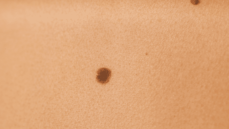
Clinical appearance
Clinically benign-looking moles can typically be removed by any appropriate technique. Clinically suspicious moles — those with ABCDE features suggesting possible melanoma — should always be excised surgically with histological analysis, never lasered or shaved. The principle: if a lesion needs definitive diagnosis, the technique must preserve the tissue for histopathology. For more on identifying suspicious features see what is the difference between a mole and a melanoma?
The patient’s skin type and scarring history
Patients with darker skin types (Fitzpatrick IV–VI) have higher rates of post-inflammatory hyperpigmentation and keloid scarring. Patients with a personal or family history of keloid formation are at higher risk. Both groups can have moles removed safely, but the technique selection, the closure method, and the post-operative scar management require additional attention. For full discussion, see do hypertrophic scars go away?
The removal techniques and when each is appropriate
Surgical excision
The most versatile and definitive technique. The mole and a small margin of surrounding skin are excised together, the wound closed with layered suturing, and the specimen sent for histology. Appropriate for:
- Any mole where definitive diagnosis is required
- Flat (intradermal) moles that cannot be addressed by shave excision
- Deep moles extending into the dermis or beyond
- Suspicious lesions
- Larger moles
- Most moles on the face where cosmetic precision matters
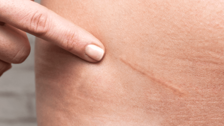
Produces a small linear scar that matures over 6–12 months. For more on what the final scar typically looks like, see what do mole removal scars look like? and how long does a mole removal scar take to fade?
Shave excision
The mole is shaved off at or just below the level of the surrounding skin with a fine blade. The wound heals as a flat mark over 2–3 weeks. Appropriate for:
- Small, raised, clinically benign moles
- Compound naevi where only the elevated component needs removal
- Patients who want a quick, simple procedure for cosmetic relief
Not appropriate for any clinically suspicious mole or any deep dermal mole. Shave excision leaves the deep component of the mole behind, which means it is not suitable when complete removal is required.
Laser mole removal
For suitable benign raised moles where histological analysis is not required, laser removal offers an alternative to surgical excision. The mole is ablated layer by layer with a precision laser. The technique is well suited to benign raised lesions on the face and other cosmetically sensitive areas. It is not appropriate for any suspicious lesion (the tissue is destroyed in the process, so no histology is possible) or for deep dermal moles.
At Centre for Surgery, the appropriate technique is chosen at consultation based on the specific lesion. Both surgical and laser options are available.
Punch excision
A small circular blade removes a cylinder of skin containing the entire mole. Used for small but deep lesions where minimising the scar matters. The wound is closed with one or two fine sutures.
When mole removal is not straightforward
A small number of cases require more careful planning:
Giant congenital melanocytic naevi
Large pigmented lesions present from birth, sometimes covering significant body areas. These have a higher lifetime melanoma risk than acquired moles and removal is often medically indicated as well as cosmetically desired. They typically require staged excision over multiple operations, sometimes with tissue expansion or skin grafting.
Multiple atypical moles
Patients with many atypical-looking moles (dysplastic naevus syndrome) need a different approach from patients with single isolated lesions. The plan typically involves careful baseline photography, regular dermoscopic surveillance, and selective excision of any lesion showing concerning change — rather than prophylactic removal of every mole.
Moles in critical anatomical locations
Moles on the eyelid margin, in the deep ear canal, near the lip vermilion, or in similar specialised areas require expertise in the anatomy of that region. Removal is safe and routine in skilled hands but should not be attempted by practitioners without the relevant training.
Moles on the soles of the feet or palms
Acral moles are more likely to be subjected to abrasion and have specific clinical features that need to be assessed dermoscopically. Acral lentiginous melanoma — the most common form of melanoma in patients with darker skin types — most often appears on these surfaces, so clinical care in assessment is important.
Moles in the genital, perianal or oral mucosal areas
Mucosal melanoma is rare but biologically aggressive, and pigmented lesions in these areas warrant careful assessment by a specialist. Removal is performed where indicated.
Can multiple moles be removed in one session?
Yes — multiple moles can usually be addressed in a single appointment, depending on:
- The total number of lesions
- Their locations (multiple moles in one area are easier to combine than scattered lesions across the body)
- The expected total local anaesthetic dose
- The expected total procedure duration
For patients with many lesions, splitting them across two or three sessions is sometimes preferable to a single long session. The plan is discussed at consultation.
What about patients on blood thinners?
Patients on antiplatelet drugs (aspirin, clopidogrel) or anticoagulants (warfarin, DOACs) can have moles removed safely, but the procedure requires adjustment. Most surgeons prefer to continue essential anticoagulation rather than stop it, and use meticulous haemostasis during the operation. Some patients may be advised to discontinue medication briefly under guidance from their prescribing doctor — but this is decided on a case-by-case basis with medical input.
It is important not to stop blood thinners without medical advice. The procedure can be planned around the medication.
What about patients with active skin conditions?
Mole removal is usually deferred until any active skin condition in the area has settled:
- Eczema or contact dermatitis in the immediate vicinity of the mole
- Acne in the area
- Active infection of the skin
- Recent significant sun damage with sunburn
This is not a contraindication to mole removal — it is a question of timing. Treating an inflamed wound bed produces worse scars than treating settled, healthy skin.
The role of histology
Every surgically excised mole at Centre for Surgery is sent for histological analysis as standard. This is critical for safety because clinical examination alone — even by experienced clinicians using dermoscopy — has an irreducible error rate. Some clinically benign-looking moles turn out on histology to be unexpectedly atypical, and a small minority turn out to be early melanoma. Histology provides the definitive cellular-level diagnosis that no clinical examination can.
For lesions removed by laser (where the tissue is destroyed in situ), no histology is available — these techniques are therefore appropriate only for clinically benign-looking lesions where the diagnostic question is already answered. For full discussion, see should every removed mole be sent for biopsy?
What we don’t recommend
- DIY mole removal at home — never safe. Risks include incomplete removal, scarring, infection, and destruction of evidence that would have identified a melanoma. See can you remove a cyst at home? for the broader case against home removal of skin lesions.
- Topical “mole removal” creams and acids sold online — unregulated, caustic, and produce worse outcomes than no treatment.
- Removal of any clinically suspicious mole by laser or shave technique — these destroy or compromise the tissue, preventing definitive histological diagnosis.
- Non-medical “mole removal” at beauty clinics — without dermoscopic assessment, without histology, and without a clinical safety net for unexpected findings.
- Skipping histology to save cost — for any surgically excised mole, histological analysis is essential to confirm what was removed.
- Mass prophylactic removal of every mole in patients with many lesions — surveillance with selective excision of changing lesions is safer and more effective than removing everything.
- Cosmetic removal without clinical assessment — every mole considered for removal should be examined first, with dermoscopy where appropriate.
Frequently asked questions
Can every mole be removed?
Almost every mole can be removed safely with the appropriate technique. The question is which technique is right for which mole, not whether removal is possible at all.
Are there moles that shouldn’t be removed?
Most can be removed if the patient wishes. Some — for example, patients with hundreds of moles where systematic surveillance is more appropriate than mass excision — are better managed by monitoring than by routine removal. Each case is assessed individually.
Is mole removal painful?
The local anaesthetic injection is the most uncomfortable part — a brief sting. The removal itself is painless. Mild soreness for 24–48 hours afterwards is normal.
Will I have a scar?
Any procedure that breaks the skin produces a scar of some kind. With plastic surgical technique on most moles, the final scar is a fine pale line that fades to barely visible over 6–12 months.
How many moles can be removed in one session?
This depends on size, location and overall local anaesthetic dose. Anywhere from one to ten or more lesions can be addressed in a single session, with the exact number assessed at consultation.
Can children have moles removed?
Yes — paediatric mole removal is offered where appropriate. Some moles are better left until the child is older; others benefit from removal sooner. We assess each case individually with the parent or guardian.
Do you offer laser mole removal?
Yes — for suitable benign raised moles where histology is not required. The choice between laser and surgical excision is made at consultation based on the specific lesion.
What happens if my mole turns out to be a melanoma?
If a privately excised mole returns a histology result showing melanoma, your surgeon will discuss this with you immediately and arrange onward management — including wider local excision if needed and referral to a specialist skin cancer multidisciplinary team.
Will my GP know about the removal?
If you would like a copy of the histology report sent to your GP for your medical record, we are happy to arrange this.
Mole removal at Centre for Surgery
Centre for Surgery is a CQC-regulated plastic surgery clinic at 95–97 Baker Street, Marylebone. Mole assessment and removal is performed by GMC-registered consultant plastic surgeons under local anaesthetic as day-case procedures. Both surgical excision with histology and laser mole removal for suitable benign moles are available. Every surgically excised specimen is sent for histological analysis as standard. No GP referral is required.
For related guides, see should every removed mole be sent for biopsy?, why choose a plastic surgeon for mole removal?, should I be concerned about an itchy or bleeding mole?, and our broader guide to common skin lumps and bumps.
Centre for Surgery · CQC-regulated · GMC specialist-registered surgeons · 95–97 Baker Street, Marylebone, London W1U 6RN · 0207 993 4849 · Book a consultation · Finance from 0% APR