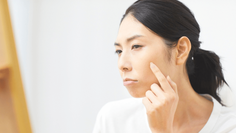

If you have noticed a lump beneath the skin and started researching it online, you have almost certainly encountered both terms: sebaceous cyst and epidermoid cyst. They are used interchangeably in most non-medical contexts — by patients, by general practice staff, and even on many healthcare websites. In clinical terms, however, they are not the same thing, and the distinction matters when it comes to understanding what you are dealing with, whether it is likely to cause problems, and what the most appropriate treatment is.
At Centre for Surgery in London, our surgeons see a high volume of patients presenting with skin cysts of all types. The majority of these are epidermoid cysts — the most common benign skin cyst in adults. A smaller number are true sebaceous cysts, which are a distinct entity with a different origin and slightly different clinical behaviour. This guide explains both conditions clearly, draws the comparison that patients are actually looking for, and explains when surgical cyst removal is the right course of action.
What Is an Epidermoid Cyst?
An epidermoid cyst — also called an epidermal inclusion cyst or, incorrectly but commonly, a sebaceous cyst — is a benign, slow-growing cyst that forms just beneath the surface of the skin. It is by far the most common type of skin cyst encountered in clinical practice.
The cyst forms when epidermal cells — the cells that make up the outermost layer of the skin — become trapped beneath the skin surface rather than shedding normally. This can happen as a result of a blocked hair follicle, minor trauma to the skin, or inflammation around a follicle. The trapped cells continue to produce keratin — the protein that forms skin, hair, and nails — which accumulates inside the cyst wall, forming a thick, cheesy, pale material that is characteristic of epidermoid cysts.
The cyst itself is lined by a stratified squamous epithelium — the same type of tissue as the outermost skin layer — and has a clearly defined wall that separates it from the surrounding tissue. This wall, known as the cyst capsule, is what makes complete surgical excision both possible and important: removing the cyst intact, including the entire capsule, is the only way to prevent recurrence.
Epidermoid cysts typically present as smooth, round, mobile swellings beneath the skin, ranging in size from a few millimetres to several centimetres. They are usually non-tender unless they become inflamed or infected. A small, central punctum — a dark spot on the skin surface — is often visible and represents the blocked follicular opening through which the cyst formed. This punctum is a helpful diagnostic feature that distinguishes epidermoid cysts from other subcutaneous lumps such as lipomas.
The most common sites for epidermoid cysts are the face, neck, scalp, back, and chest — areas with a high density of hair follicles. They can, however, develop anywhere on the body. They are more common in adults than in children and show a slight male predominance.
RELATED: Will a Cyst Come Back After Removal?
What Is a True Sebaceous Cyst?
A true sebaceous cyst is considerably rarer than an epidermoid cyst and has a genuinely different origin. True sebaceous cysts arise from the sebaceous glands themselves — the oil-producing glands attached to hair follicles — rather than from trapped epidermal cells. They are lined by glandular epithelium rather than squamous epithelium, and they contain sebum — the oily secretion produced by sebaceous glands — rather than keratin.
The two most clinically relevant examples of true sebaceous cysts are trichilemmal cysts (also called pilar cysts) and steatocystoma multiplex. Trichilemmal cysts are the more common of the two and are particularly associated with the scalp — they account for the majority of cysts encountered in the scalp and have a characteristic thick, white, odourless content and a smooth, firm wall. They have a hereditary component, often running in families, and can be multiple. Steatocystoma multiplex is a rare condition in which numerous small sebaceous cysts develop across the body, with a predilection for the trunk, axillae, and upper arms.
In practice, true sebaceous cysts are far less frequently encountered than epidermoid cysts. When a general practitioner or patient refers to a “sebaceous cyst” in everyday clinical conversation, they are almost always describing what is technically an epidermoid cyst. This is not a significant problem in most clinical contexts — both are benign, both are managed similarly, and the distinction rarely changes the treatment approach. Where it does matter is in the pathological analysis of removed tissue, which is why tissue from cyst excisions is routinely sent for histological examination at Centre for Surgery.
Epidermoid Cyst vs Sebaceous Cyst — The Key Differences
Origin
Epidermoid cysts originate from trapped epidermal cells, typically at a blocked hair follicle or a site of minor skin trauma. True sebaceous cysts originate from the sebaceous glands themselves and are lined by glandular rather than squamous epithelium.
Content
Epidermoid cysts contain keratin — a thick, pale, cheesy material that often has a characteristic odour when the cyst is opened or ruptures. True sebaceous cysts contain sebum, which is oily and has a different consistency. In practice, the content can help identify the cyst type during surgical excision, though histological examination of the cyst wall provides definitive classification.
Location
Epidermoid cysts can occur anywhere on the body but are most common on the face, neck, trunk, and back. Pilar cysts — the most common true sebaceous cysts — have a very strong predilection for the scalp. If a patient presents with a cyst on the scalp, a pilar cyst is considerably more likely than an epidermoid cyst in that location.
Punctum
The presence of a central punctum on the skin surface is characteristic of epidermoid cysts. Pilar cysts typically do not have a visible punctum, which contributes to their smooth, featureless surface appearance. The absence of a punctum on a scalp cyst is a useful clinical pointer towards a pilar rather than epidermoid origin.
Hereditary Tendency
Pilar cysts have a recognised hereditary component — they frequently run in families and often present as multiple cysts rather than a solitary lesion. Epidermoid cysts are generally sporadic, though some are associated with conditions such as Gardner syndrome when multiple lesions develop across the body.
Malignant Potential
Both epidermoid and pilar cysts are overwhelmingly benign. Very rarely, malignant transformation can occur in either — epidermoid cysts can very occasionally give rise to squamous cell carcinoma, and pilar cysts can rarely develop into a malignant pilar tumour (also called a proliferating trichilemmal cyst). These events are genuinely rare, but they underscore the value of sending excised tissue for histological analysis rather than discarding it, which is standard practice at Centre for Surgery.
How Are These Cysts Diagnosed?
In most cases, both epidermoid and pilar cysts can be diagnosed clinically — on the basis of the history and physical examination alone. A smooth, round, mobile, non-tender swelling beneath the skin, with or without a central punctum, in a typical anatomical location, has a very high probability of being one of these two benign cyst types.
Where clinical examination leaves doubt — for example, if the lump is deeper than expected, if it is rapidly growing, if it is hard rather than soft, or if there is clinical suspicion of an alternative diagnosis — ultrasound imaging can be a helpful adjunct. Ultrasound can characterise the cyst’s structure, confirm its relationship to surrounding tissues, and identify features that might suggest a different diagnosis such as a lipoma, lymph node, or vascular lesion. In cases where the diagnosis is genuinely uncertain, further imaging with MRI may occasionally be required before surgical planning.
Definitive diagnosis is established by histological examination of the excised specimen. At Centre for Surgery, all tissue removed during lumps and bumps removal procedures is routinely sent for pathological analysis. This is not merely a clinical formality — it provides the patient with the reassurance of a confirmed benign diagnosis, and it ensures that any unexpected finding is identified promptly and managed appropriately.
RELATED: Should Every Removed Mole Be Sent for Biopsy?
When Do Cysts Cause Problems?
Many epidermoid and pilar cysts remain entirely asymptomatic for years and never require treatment. Patients often describe a cyst that has been present for a long time without causing any discomfort, which has recently begun to trouble them either because it has grown or because it has become inflamed.
Inflammation and Infection
The most common complication of both cyst types is inflammation, which can progress to infection. When the cyst wall breaks down — either spontaneously or following minor trauma — the keratin or sebum contents provoke a strong inflammatory response in the surrounding tissue. The cyst becomes red, hot, swollen, and very tender, and a surrounding area of erythema and induration may develop rapidly. In more severe cases, the inflammatory response can progress to frank abscess formation, with the accumulation of pus in and around the cyst cavity.
An inflamed or infected cyst is not suitable for immediate surgical excision — the inflamed tissue planes make clean dissection difficult, increase the risk of incomplete removal, and significantly raise the chance of wound complications. The standard management is to treat the acute inflammation first — with oral antibiotics if infection is confirmed, with incision and drainage if an abscess has formed — and to plan definitive excision once the inflammation has fully settled, typically four to eight weeks later.
This is one of the most important points to convey to patients: presenting for cyst removal when the cyst is calm, non-inflamed, and clearly defined is always better than waiting until an inflammatory episode forces the issue. Elective excision of a quiescent cyst is a cleaner, more straightforward procedure with a lower risk of incomplete removal and a better cosmetic outcome than emergency drainage or excision of an acutely inflamed cyst.
Size and Cosmetic Concern
Cysts that are enlarging, that have reached a size that causes physical discomfort from pressure on surrounding structures, or that are in a visible location causing cosmetic concern are all appropriate candidates for elective surgical removal. There is no minimum or maximum size threshold — the decision to remove is based on the patient’s individual circumstances, the clinical assessment, and the patient’s own preferences and goals.
Location-Specific Problems
Cysts in certain locations can cause specific functional problems. Scalp cysts that are large or multiple can create difficulty with hair styling and grooming. Cysts on the face — particularly near the eyes, on the nose, or along the jawline — can be cosmetically prominent and difficult to conceal. Cysts in pressure areas such as the back of the neck or the buttocks can cause discomfort when sitting or lying down. All of these represent reasonable indications for removal.
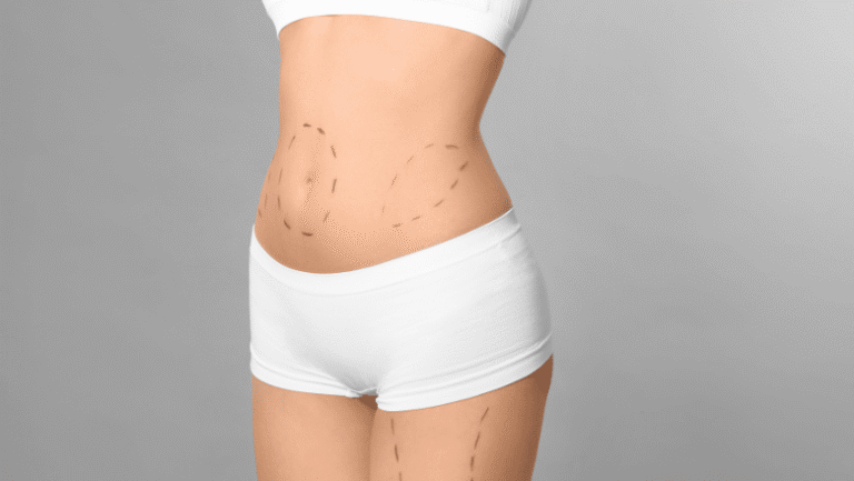
How Are Epidermoid and Sebaceous Cysts Removed?
Surgical excision is the definitive treatment for both cyst types. The procedure is typically performed under local anaesthesia as a day case at Centre for Surgery’s Baker Street clinic and takes between 20 and 45 minutes depending on the size and location of the cyst.
Minimal Excision Technique
For smaller, uncomplicated cysts, the minimal excision technique is often used. A small incision — typically 2 to 4mm — is made through the skin over the cyst, and the cyst contents are expressed. The collapsed cyst wall is then carefully removed through the small opening using fine forceps. The advantage of this approach is the minimal scarring produced by the tiny incision. The technique is most effective when the cyst wall is intact and the cyst has not been previously inflamed or infected.
Standard Elliptical Excision
For larger cysts, cysts with a prominent punctum, or those with a history of previous inflammation where scarring may have made the wall more adherent, standard elliptical excision is performed. An ellipse of skin encompassing the punctum is excised together with the cyst, ensuring the entire cyst capsule is removed en bloc. The wound is closed with fine sutures, which are removed at a follow-up appointment. This technique provides the highest certainty of complete cyst removal and is the standard approach for most facial and neck cysts where the cosmetic result is a priority.
Complete Capsule Removal — Why It Matters
Regardless of technique, complete removal of the cyst capsule is essential. If any portion of the capsule is left behind, the residual lining tissue will continue to produce keratin or sebum, and the cyst will reform — often within months. Recurrence following incomplete excision is the most common reason patients present for repeat cyst surgery. The importance of complete excision is also why attempting to drain or squeeze a cyst at home — without removing the capsule — invariably results in the cyst returning.
Can Cysts Be Removed on the NHS?
Removal of benign skin cysts is not routinely available on the NHS in most parts of England. NHS guidelines classify excision of asymptomatic or cosmetically bothersome cysts as a procedure of low clinical priority, meaning that referral and funding for the procedure through the NHS is unlikely to be granted unless the cyst is causing significant functional impairment or there is clinical concern about the diagnosis. The same policy that restricts access to NHS mole removal applies broadly to cyst removal and other benign skin lesion procedures.
Patients who require cyst removal and are unable to access it through the NHS should seek treatment privately. Centre for Surgery provides a straightforward, consultant-led pathway for cyst removal at our Baker Street clinic, with transparent pricing, no waiting lists, and a comprehensive aftercare package. Finance options including 0% APR through Chrysalis Finance are available for patients who prefer to spread the cost.
Recovery After Cyst Removal
Recovery from cyst excision is straightforward in the vast majority of cases. The local anaesthetic injection is the most uncomfortable part of the procedure for most patients — the excision itself is painless. Following surgery, the area will be mildly tender for two to three days, and there may be some minor bruising and swelling around the wound site. Most patients find that standard over-the-counter pain relief such as paracetamol is entirely adequate for any post-operative discomfort.
The wound should be kept clean and dry for the first 48 hours. Sutures, where placed, are typically removed at five to seven days for facial wounds and seven to ten days for body wounds. Most patients return to their normal activities — including office-based work — the day after surgery. Strenuous physical activity involving the operated area should be avoided for one to two weeks.
The final scar appearance continues to improve over six to twelve months as the healed tissue matures. In the majority of cases, the scar from a well-performed cyst excision is a fine, pale line that is considerably less noticeable than the cyst it replaced — particularly for facial and neck cysts where patients have often been self-conscious about the visible lump for some time before seeking treatment.
RELATED: Earlobe Cyst — How to Get Rid of a Lump in the Earlobe
Frequently Asked Questions
Is a sebaceous cyst the same as an epidermoid cyst?
Not technically, though the terms are used interchangeably in most contexts. A true sebaceous cyst arises from the sebaceous gland and is lined by glandular epithelium. An epidermoid cyst arises from trapped skin cells and is lined by squamous epithelium. In practice, the vast majority of cysts referred to as sebaceous cysts are epidermoid cysts. Both are benign and managed similarly.
How do I know if my cyst is infected?
Signs of a cyst becoming infected include rapid increase in size, redness and warmth of the overlying skin, increasing tenderness, fluctuance (a feeling of fluid under tension), and occasionally discharge of pus through the skin surface or punctum. A fever may also be present in more significant infections. If you notice these signs, contact your GP or surgical team promptly.
Can I squeeze or drain a cyst myself?
No. Squeezing or attempting to drain a cyst at home will not resolve it — the cyst capsule remains in place and the cyst will refill. Squeezing can also introduce bacteria into the cyst, triggering infection and inflammation that significantly complicates subsequent surgical removal. Full guidance on why this is inadvisable is covered in our article on home cyst removal.
Will my cyst grow back after removal?
Not if the entire cyst capsule is removed at surgery. Recurrence following complete excision is uncommon. Recurrence following incomplete removal — which is more likely after drainage procedures, inflamed excisions, or attempted home removal — is common. Choosing an experienced surgeon who prioritises complete capsule excision is the most important factor in preventing recurrence. More detail is covered in our guide to whether cysts come back after removal.
Does cyst removal leave a scar?
Yes — any incision produces a scar. However, the scar from a well-performed cyst excision using fine sutures and meticulous technique is typically a fine, pale line that is far less noticeable than the cyst itself. Scars continue to mature and improve for up to twelve months following surgery.
How much does cyst removal cost at Centre for Surgery?
The cost of cyst removal at Centre for Surgery depends on the size, location, and complexity of the cyst. Pricing is provided at your consultation following clinical assessment. Finance options including 0% APR through Chrysalis Finance are available.
Cyst Removal at Centre for Surgery
Mr Andreas Shiatis is a consultant plastic surgeon at Centre for Surgery with extensive experience in the surgical management of skin cysts and lumps and bumps removal. He trained on the London Deanery plastic surgery rotation, holds the FRCS(Plast) from the Royal College of Surgeons of England, and is on the GMC specialist register for plastic surgery. His approach to cyst removal prioritises complete capsule excision, precise wound closure, and a cosmetic outcome that is as discreet as possible.
All cyst excision procedures at Centre for Surgery are performed at our CQC-regulated Baker Street clinic in central London. Every excised specimen is sent for histological analysis as standard. Finance options including 0% APR through Chrysalis Finance are available — visit our Finance Options page for details.
Phone: 0207 993 4849 | Email: contact@centreforsurgery.com | Address: 95-97 Baker Street, London W1U 6RN

[contact-form-7 id=”256″ title=”Treatments form”]