Hyperpigmentation is the visible darkening of patches of skin caused by overproduction or uneven distribution of melanin — the pigment that gives skin its colour. It affects people of all skin types but disproportionately affects Fitzpatrick types IV to VI, where it’s more common, more persistent and more difficult to treat. Understanding the specific cause matters because the wrong treatment can make the problem worse — particularly with melasma, where heat-based interventions often trigger a flare.
This guide explains the four main types of hyperpigmentation, what drives each, and which treatment is appropriate for which type. At Centre for Surgery, our laser specialists treat laser pigmentation using the Fotona SP Dynamis Pro at our CQC-regulated Baker Street private hospital, with protocols matched to type and Fitzpatrick skin tone.
How hyperpigmentation forms
Pigment production happens in melanocytes — specialised cells in the basal layer of the epidermis. Melanocytes synthesise melanin and transfer it to surrounding keratinocytes via small packets called melanosomes. Normal skin colour reflects the balance of melanin production, distribution and turnover.
Hyperpigmentation develops when this balance is disrupted in one or more ways:
- Increased melanin production — melanocytes are stimulated to make more pigment than the skin needs
- Abnormal distribution — melanin clusters unevenly rather than spreading uniformly
- Slower clearance — pigment-containing cells aren’t shed at the normal rate, so pigment accumulates
- Deeper deposition — pigment ends up in the dermis as well as the epidermis, where it’s significantly harder to treat
Different triggers drive different patterns. The four main clinical types — sunspots, melasma, post-inflammatory hyperpigmentation and drug-induced pigmentation — each have distinct mechanisms and respond to different treatments.
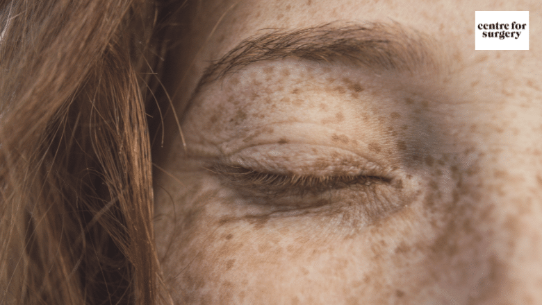
Sunspots (solar lentigines)
Sunspots — also called age spots, liver spots or solar lentigines — develop after years of cumulative UV exposure. UV radiation triggers melanocytes to produce more melanin as a defensive response. Where exposure is uneven across the skin, melanin clusters form discrete brown patches.
The clinical features:
- Flat, well-defined brown or tan patches
- Usually round or oval with clear borders
- Vary in size from a few millimetres to over a centimetre
- Appear on sun-exposed areas: face, backs of hands, forearms, upper chest, shoulders
- Develop gradually over years; more common after age 40 in fair skin, earlier in darker skin
The pigment in sunspots sits relatively superficially in the epidermis, which makes them one of the more treatable forms of hyperpigmentation. They respond well to ablative laser resurfacing (Erbium YAG laser), Q-switched lasers, IPL and chemical peels. Topical retinoids, vitamin C and azelaic acid can fade superficial spots over months but are slower than in-clinic interventions.
Suspicious changes — irregular borders, varied colouration within a single spot, sudden growth, bleeding — should be assessed before any cosmetic treatment, as they can indicate skin lesions needing diagnostic evaluation rather than aesthetic clearance.
Melasma
Melasma presents as larger, symmetrical patches typically across the cheeks, forehead, upper lip, chin and nasal bridge. It’s driven by a combination of hormonal influence, UV exposure and heat — which is why it often appears or worsens during pregnancy, with the combined oral contraceptive pill, or with hormone replacement therapy.
Clinical features:
- Diffuse, blotchy patches with indistinct edges (unlike the well-defined borders of sunspots)
- Symmetrical distribution — both cheeks, both sides of the upper lip
- Light brown to dark brown, sometimes with a greyish tint when dermal involvement is present
- Predominantly affects women between 20 and 40, particularly Fitzpatrick types III to VI
- Often worsens during pregnancy (“chloasma” or “pregnancy mask”) and improves postpartum
- Significantly worsens with UV exposure and visible light
Unlike sunspots, the pigment in melasma can sit at multiple depths in the skin, including deep dermal pigment that no topical treatment can reach. The mixed epidermal-dermal pattern is what makes melasma so stubborn to treat — and why heat-based treatments such as IPL or aggressive laser resurfacing can backfire, stimulating more melanin production and worsening the condition.
The right approach for melasma:
- Daily SPF 50 with iron oxide content (which blocks visible light as well as UV)
- Prescription topicals — hydroquinone, tretinoin, azelaic acid, often as a triple combination (“modified Kligman formula”)
- Low-energy laser modalities such as low-fluence Q-switched Nd:YAG
- Dermamelan or similar specialist peels — see our guide on the Dermamelan peel
- Tranexamic acid (oral or topical) for moderate to severe cases under specialist supervision
Avoid heat-based treatments. Treatment is long-term — melasma is managed rather than cured. For a fuller treatment overview, see our companion guide on melasma causes and solutions.
Post-inflammatory hyperpigmentation (PIH)
PIH is the dark mark left after any inflammatory event in the skin. The most common causes are acne, eczema, an injury, an insect bite, or an aggressive cosmetic procedure performed at the wrong settings for the skin type. PIH is significantly more common and more persistent in skin types IV–VI on the Fitzpatrick scale.
The mechanism:
- Skin inflammation stimulates melanocytes to overproduce melanin
- Some of the excess melanin is deposited deeper in the skin, into the dermis
- Once the inflammation settles, the pigment remains
- Resolution depends on the body clearing the pigment — a slow process, particularly for dermal deposits
Clinical features:
- Brown or darkened patches in areas where skin was previously inflamed
- Pattern follows the original inflammation — round/oval if from acne lesions, linear if from scratches
- Lighter PIH (epidermal) is brown; darker PIH (dermal) can appear greyish-blue
- Some fades spontaneously over months; some persists for years
For superficial PIH, topical retinoids, vitamin C, azelaic acid and gentle chemical peels are effective over several months. For deeper or more persistent PIH, non-ablative fractional laser can help. Caution is essential in darker skin types — overly aggressive treatment can drive a paradoxical worsening.
The most important step in PIH management is preventing further inflammation. Active acne should be controlled — see our hub guide on the most effective treatment for acne — and ongoing trigger control is essential, including strict sun protection.
Drug-induced pigmentation
A handful of medications cause skin darkening, either through direct pigment deposition or by sensitising the skin to UV. The common culprits:
- Antibiotics — particularly minocycline, doxycycline (less commonly)
- Antimalarials — hydroxychloroquine, chloroquine
- Chemotherapy agents — busulfan, bleomycin, cyclophosphamide
- Amiodarone — cardiac antiarrhythmic, causes a characteristic blue-grey pigmentation
- Some psychotropic medications — chlorpromazine and other phenothiazines
- Heavy metals — silver, gold, iron containing preparations
If pigmentation appears suddenly without an obvious cause, the patient’s medication list is worth reviewing. Drug-induced pigmentation often fades after the medication is stopped, though some forms — particularly amiodarone-related — can persist for years.
Treatment depends on the specific drug and pattern. Stopping or substituting the responsible medication (where clinically possible) is the first step. Laser treatment can help in some cases but isn’t universally effective for all drug-induced patterns.
Other contributors to hyperpigmentation
Beyond the four main types, several other factors contribute to or worsen pigmentation:
Genetic factors
The tendency to develop hyperpigmentation runs in families. Fitzpatrick skin type is the dominant genetic factor, but family-specific patterns also matter. Patients with first-degree relatives who developed melasma during pregnancy are themselves at higher risk.
Age
Cumulative UV damage over decades drives age-related sunspots. The number of melanocytes decreases with age, but the remaining cells often become hyperactive in specific clusters, producing the characteristic age-spot pattern.
Hormonal changes
Beyond pregnancy and the combined oral contraceptive pill, perimenopause and HRT can trigger or worsen melasma. PCOS and other endocrine conditions also contribute. Hormonal changes don’t directly cause hyperpigmentation, but they sensitise melanocytes to other triggers.
Friction and pressure
Chronic friction (tight clothing, persistent rubbing) can drive friction-related hyperpigmentation, particularly in flexural areas and on darker skin types.
Phototoxic reactions
Some plants (parsnip, citrus oils, fig sap) and chemicals contain compounds that cause exaggerated skin reaction to UV light. The classic example is “berloque dermatitis” from bergamot oil in perfume — a streaky hyperpigmentation pattern on the neck.
Skin conditions
Several inflammatory skin conditions — lichen planus, atopic dermatitis, lupus, contact dermatitis — can leave significant PIH, especially in darker skin types. Treating the underlying condition is the first step.
Why correct diagnosis matters
The most common mistake — made by patients self-treating, and by less experienced practitioners — is treating melasma as if it were sunspots. The aggressive lasers and IPL settings that fade sunspots in one or two sessions can drive melasma into a worse flare that takes months to settle.
A proper consultation should establish:
- Which type of hyperpigmentation is present
- Where in the skin the pigment sits (epidermal, dermal, or mixed)
- What triggers are still active (sun, hormones, ongoing inflammation, medications)
- What treatments are appropriate for the type, the depth, and the skin’s Fitzpatrick category
- What can be expected — clearance, partial improvement, or maintenance control
The depth of pigment is often assessed using a Wood’s lamp (which makes superficial epidermal pigmentation appear darker but doesn’t affect dermal pigment) or by applying mild skin tension (epidermal pigment lightens as skin stretches, dermal pigment doesn’t change). These simple tests guide treatment choice.
Treatment by type — a quick reference
| Type | First-line treatment | Avoid |
|---|---|---|
| Sunspots | Ablative laser, Q-switched laser, IPL, chemical peels | Nothing specific — most treatments work |
| Melasma | SPF 50 + topicals (hydroquinone, tretinoin, azelaic acid), low-fluence Q-switched Nd:YAG, Dermamelan peel | Heat-based treatments, IPL, aggressive ablative laser |
| PIH (superficial) | Topical retinoids, vitamin C, azelaic acid, gentle peels | Aggressive resurfacing in darker skin types |
| PIH (deep) | Non-ablative fractional laser, careful Q-switched protocols | High-fluence settings without patch testing |
| Drug-induced | Stop responsible drug if possible; laser case-by-case | Aggressive treatment before drug substitution |
For more on melasma specifically, see melasma hyperpigmentation — causes and solutions. For sun damage more broadly, see our guide on the five signs of sun damage.
Prevention
Once hyperpigmentation has developed, prevention is the most important step in stopping progression and preserving any treatment result. The non-negotiables:
- Daily broad-spectrum SPF 50 — every day, including overcast days and indoors near windows. For melasma specifically, choose products containing iron oxide for visible light protection.
- Physical sun protection — wide-brimmed hat, UV-protective sunglasses, avoid peak hours (10am to 4pm in summer)
- Avoid tanning — both natural and tanning beds
- Gentle skincare — avoid harsh exfoliants and aggressive actives that drive inflammation in pigment-prone skin
- Treat triggers — control active acne, manage hormonal influences where possible, review medications with your GP if drug-induced pigmentation is suspected
- Hands-off — don’t pick at lesions; don’t squeeze blemishes. Both worsen PIH risk.
What we don’t recommend
- Aggressive at-home brightening protocols — high-percentage hydroquinone, unregulated “skin lightening creams” from outside the UK regulatory framework, or DIY peels can drive inflammatory hyperpigmentation that’s worse than the original problem
- IPL or aggressive laser for melasma — heat-based treatments often worsen melasma. Recognise the type before treating.
- Microdermabrasion for established pigmentation — superficial mechanical abrasion doesn’t reach where the pigment lives and can drive inflammation. Not part of our offering.
- Treatment without sun protection commitment — there’s no point treating pigmentation without addressing ongoing UV exposure. The result will simply regenerate.
- Quick-fix promises — some pigmentation patterns (particularly melasma and deep PIH) genuinely need long-term management. Anyone promising one-session clearance is overpromising.
Frequently asked questions
Can hyperpigmentation be permanently removed?
For sunspots, yes — single laser sessions often produce permanent clearance of individual spots, with new spots developing only with new sun exposure. For melasma, the answer is no — melasma is managed rather than cured. For PIH, most patients achieve clearance over time with proper treatment but the underlying tendency to develop PIH from new inflammation persists.
How long until I see results?
Laser treatment of sunspots: visible improvement within 2 to 3 weeks. Topical regimens: 8 to 12 weeks for visible benefit. Chemical peels: 2 to 4 weeks. Melasma management: ongoing, with maintenance rather than clearance as the goal.
Is laser safe for darker skin types?
Yes, with proper protocol calibration. Nd:YAG at 1,064 nm is the safest wavelength for Fitzpatrick types IV–VI. Patch testing is recommended for V–VI; conservative initial settings; extended sun avoidance pre- and post-treatment.
Will my hyperpigmentation come back?
Depends on type and ongoing trigger control. Sunspots can return with new UV exposure. Melasma reliably returns without ongoing management. PIH can develop from any new inflammation in the same area.
Can I treat hyperpigmentation while pregnant?
Treatment options are restricted during pregnancy. Most laser and topical retinoid treatments are avoided. Mineral SPF, gentle skincare and avoiding triggers are the safe pregnancy approach. Active treatment can resume after pregnancy and breastfeeding.
What’s the difference between sunspots and freckles?
Freckles (ephelides) typically develop in childhood, are smaller, fade in winter, and are common in fair, often red-haired patients. Sunspots develop in adulthood from cumulative UV damage, are larger, don’t fade in winter, and are present in all skin types but more common in older fair skin.
How much does treatment cost?
Pricing depends on the type and area treated. Single-spot laser treatment starts from modest single-session pricing; full-face pigmentation programmes for melasma involve longer commitments. A consultation gives an exact quote. Finance from 0% APR is available through Chrysalis Finance.
Why choose Centre for Surgery
Our laser specialists treat hyperpigmentation on the Fotona SP Dynamis Pro at our CQC-regulated Baker Street private hospital. Every consultation establishes the type, the depth, the active triggers and the Fitzpatrick category before treatment is recommended — because the wrong treatment can make pigmentation worse. The right treatment, calibrated to your skin, achieves the results that aggressive over-treatment can’t.
Centre for Surgery · CQC-regulated · GMC specialist-registered surgeons · 95–97 Baker Street, Marylebone, London W1U 6RN · 0207 993 4849 · Book a consultation · Finance from 0% APR