If you have been told one of your skin growths is a seborrhoeic keratosis — or you have looked one up and landed on the word “melanoma” — the question that follows is understandable and important: can a seborrhoeic keratosis turn into melanoma? The short, reassuring answer is no. A seborrhoeic keratosis is a benign growth, and it does not transform into skin cancer. But there is an important nuance behind that simple answer, and it is the reason this question matters so much: melanoma can occasionally look like a seborrhoeic keratosis, and in rare cases the two can sit side by side. Understanding the difference — and knowing which signs should prompt a professional check — is genuinely worth a few minutes of your time.
This guide explains what a seborrhoeic keratosis is, why it cannot become melanoma, how to tell the two apart, and — most importantly — the specific warning signs that mean a growth should be assessed by a specialist rather than assumed to be harmless. At Centre for Surgery, this assessment sits within our wider skin lesion removal service, and benign lesions confirmed at consultation can be treated through our seborrhoeic keratosis removal service — while any growth with worrying features is examined first and directed to the right pathway, including onward referral for specialist skin cancer care where it is needed.
The short answer: no, but here is the nuance
A seborrhoeic keratosis cannot turn into a melanoma. The two are fundamentally different types of growth. A seborrhoeic keratosis develops from keratinocytes — the ordinary cells that make up the outer layer of the skin. Melanoma develops from melanocytes — the pigment-producing cells. Because they arise from different cell types, one does not convert into the other. A seborrhoeic keratosis you have had for years is not quietly becoming dangerous.
So why does the question come up so often? Because of appearances. Seborrhoeic keratoses are frequently pigmented — tan, dark brown, or even close to black — and that dark colour is exactly what makes people worry about melanoma. On top of that, there are two genuine clinical situations worth being aware of: first, a melanoma can occasionally mimic a seborrhoeic keratosis closely enough to be mistaken for one; and second, in rare cases a melanoma can develop in the skin immediately next to, or even within the same area as, a seborrhoeic keratosis. Neither of these means the keratosis “became” cancer — but both are excellent reasons not to simply assume a dark growth is harmless without a proper look.
What is a seborrhoeic keratosis?
A seborrhoeic keratosis (sometimes called a seborrhoeic wart, or by the older term “senile wart”) is one of the most common benign skin growths in adults. Most people over 50 have at least a few, and many have dozens. They tend to increase in number with age, and there is a strong hereditary tendency — if your parents had a lot of them, you probably will too.
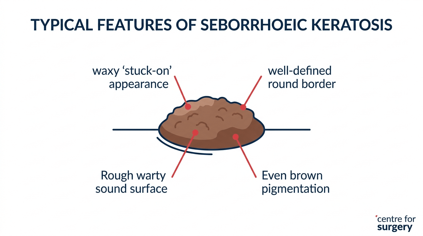
The classic features are quite recognisable once you know them. A typical seborrhoeic keratosis has a waxy, “stuck-on” appearance, as though a small piece of candle wax or a barnacle has been placed on the skin surface rather than growing out of it. The surface is often rough or warty, the border is usually well-defined and rounded, and the colour — though it can be dark — is generally even across the whole growth. They commonly appear on the trunk, back, chest, face and scalp. They are entirely harmless, but people often choose to have them removed because they catch on clothing and jewellery, become irritated, or are simply unwelcome cosmetically — particularly on the face, where lesion removal calls for techniques chosen to minimise scarring in cosmetically sensitive areas.
Crucially, a seborrhoeic keratosis is benign from the outset and stays benign. Removal, when people choose it, is for comfort or appearance — not because the growth poses any health risk.
What is melanoma, and how is it different?
Melanoma is a serious form of skin cancer that arises from melanocytes, the cells that give skin its pigment. Unlike a seborrhoeic keratosis, melanoma is malignant: it can grow into deeper tissue and, if not caught early, spread elsewhere in the body — which is why confirmed cases are managed through dedicated skin cancer pathways rather than cosmetic treatment. It is strongly linked to ultraviolet exposure — sun and sunbeds — and it can appear either within an existing mole or as a brand-new mark on otherwise normal skin.
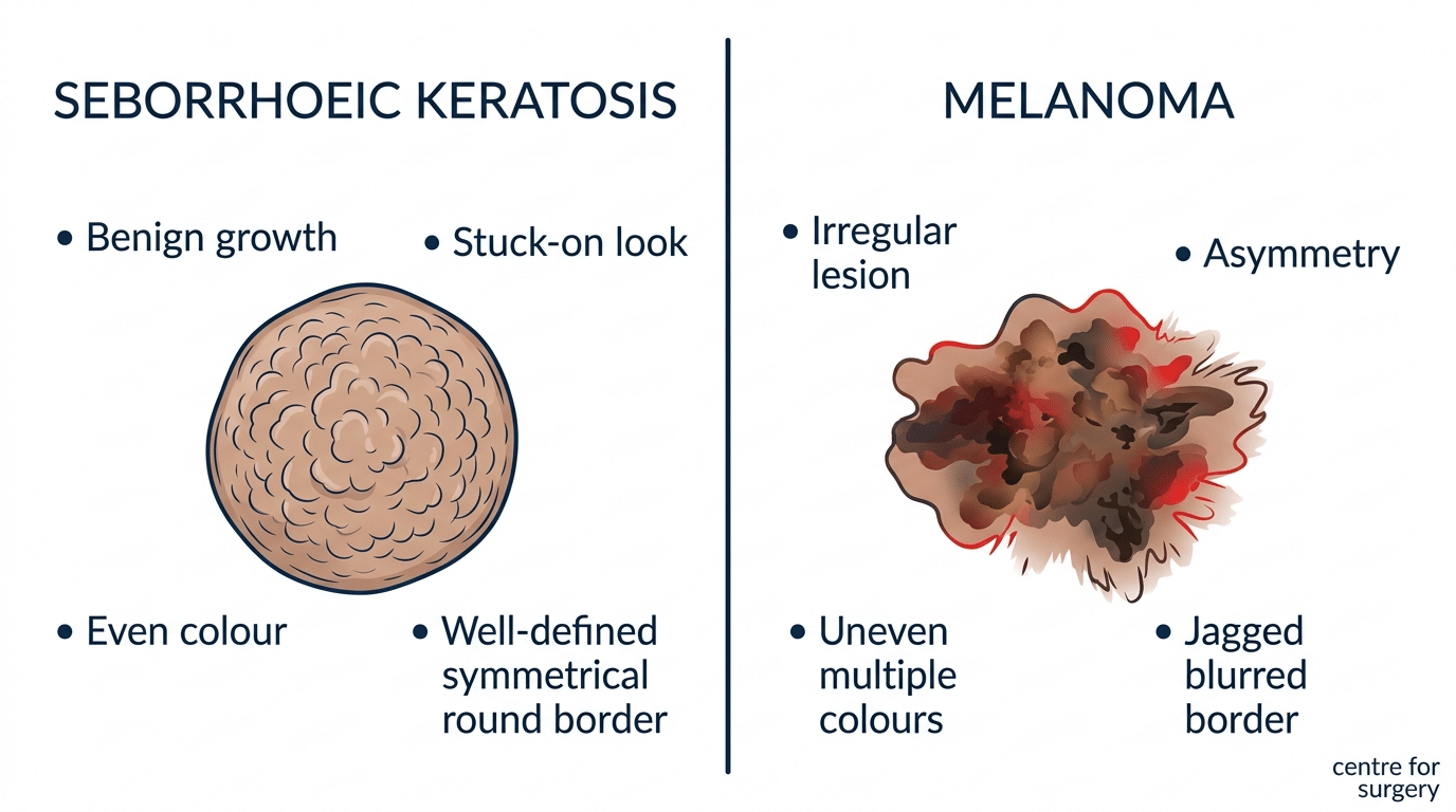
The visual differences, in most cases, are meaningful. Where a seborrhoeic keratosis tends to be symmetrical, evenly coloured, and neatly bordered with that stuck-on look, a melanoma more often shows asymmetry, an uneven mix of colours, and an irregular, blurred or jagged edge. A seborrhoeic keratosis usually sits on the skin surface; a melanoma tends to look and feel more integrated into the skin. Where several seborrhoeic keratoses often appear together and look similar to one another, a melanoma is typically a solitary lesion that stands out from the person’s other marks — what dermatologists sometimes call the “ugly duckling” sign. This is also why a single, new or changing mole that looks different from your others deserves particular attention.
That said — and this is the heart of why the question is worth taking seriously — these distinctions are not always obvious to the naked eye. Some melanomas are very good mimics of benign growths. This is precisely why professional assessment, often with a dermatoscope, exists.
Why melanoma can be mistaken for a seborrhoeic keratosis
If seborrhoeic keratoses and melanomas are so different underneath, why do they get confused at all? The answer is that appearances overlap more than most people expect. Both can be dark brown or black. Both can have a slightly raised, textured surface. And some melanomas — particularly a variety that produces a thickened, warty surface — can take on a genuinely “keratosis-like” look that even experienced clinicians treat with caution. Medical literature documents cases where a growth confidently assumed to be a benign seborrhoeic keratosis turned out, on laboratory examination, to be a melanoma in disguise.
There is a second scenario worth understanding. Because seborrhoeic keratoses are so common in older skin, and melanoma risk also rises with age and sun exposure, it is entirely possible for a melanoma to develop on skin that is already dotted with benign keratoses. A new or changing dark spot can therefore be hiding in plain sight among a scatter of harmless growths, which is exactly why the “odd one out” — the lesion that does not match its neighbours — deserves particular attention. None of this means your keratoses are dangerous. It means that “it is probably just another seborrhoeic keratosis” is a reasonable everyday assumption but a poor substitute for a proper look when a growth is behaving differently from the rest.
This is also why reputable clinics do not simply destroy every pigmented growth on request without assessing it first. Removing a lesion with a method that leaves no tissue behind — before confirming what it is — would discard the very evidence needed to catch a melanoma early. Diagnosis comes first; removal method follows from it.
The ABCDE warning signs
A widely used checklist for spotting a potentially concerning pigmented lesion is the ABCDE rule. It is a helpful memory aid for the general public, though it is not a diagnosis — it is a prompt to get something looked at.
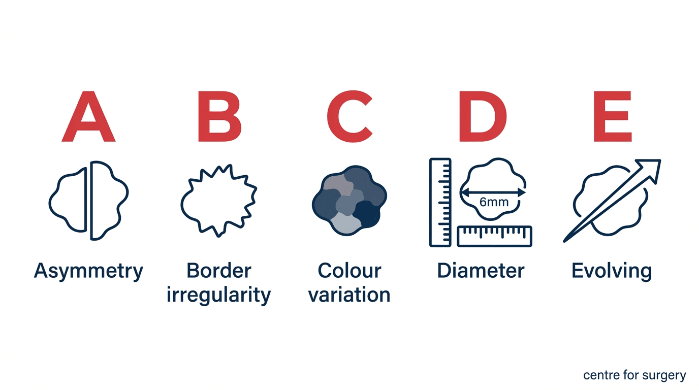
A — Asymmetry. One half of the lesion does not match the other half.
B — Border. The edges are irregular, ragged, notched or blurred rather than smooth and well-defined.
C — Colour. The colour is not uniform — there may be several shades of brown, black, and sometimes patches of red, white or blue.
D — Diameter. The lesion is larger than about 6mm (roughly the size of a pencil eraser), although melanomas can be smaller.
E — Evolving. The lesion is changing — in size, shape, colour, or elevation — or it has begun to bleed, itch or crust. Change over time is one of the most significant warning signs of all.
If a growth you have been thinking of as a seborrhoeic keratosis shows any of these features, do not rely on the assumption that it is benign. Have it assessed.
When should a seborrhoeic keratosis be checked?
Most seborrhoeic keratoses never need medical attention at all. But because a small number of melanomas can masquerade as benign growths, there are specific circumstances where a professional check is the sensible course of action. The guide below shows what is reassuring and what should prompt an assessment.
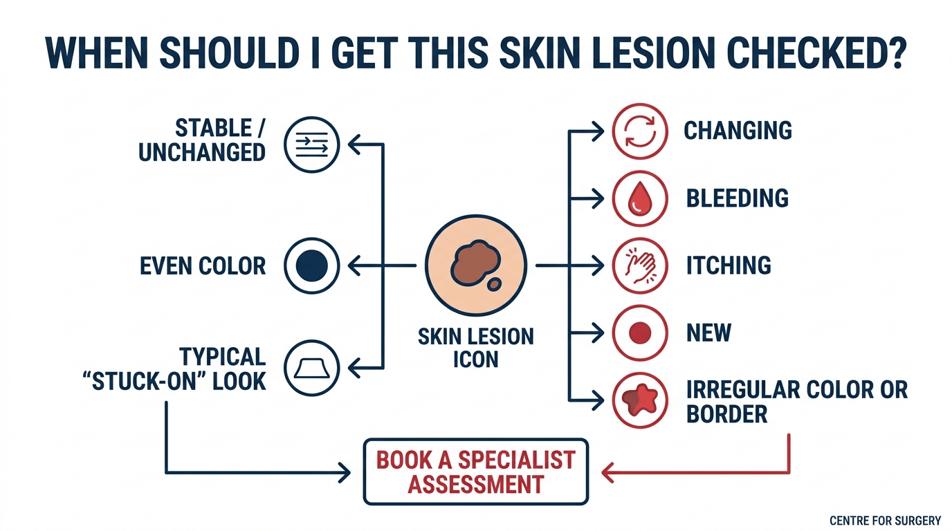
It is reasonable to simply monitor a growth that has been stable for years, has an even colour, a well-defined border, and the typical stuck-on look of a seborrhoeic keratosis — particularly when it looks similar to several others you have. On the other hand, you should arrange a specialist assessment if a growth is new and different from your others, is changing in size, shape or colour, has started to bleed, itch or become crusted without obvious injury, has an irregular border or an uneven mix of colours, or simply stands out as the odd one out among your other marks. When in doubt, the safe and simple step is to have it examined — a benign growth confirmed as benign costs you nothing but a little time, whereas an early melanoma caught promptly is far more treatable. If you are weighing up your options, our guide on whether you can get mole removal on the NHS explains how NHS and private assessment differ for growths like these.
Other growths confused with a seborrhoeic keratosis
Melanoma is the most important lesion to distinguish from a seborrhoeic keratosis, but it is not the only one people mix up. Ordinary moles can look similar, especially when raised and pigmented. Viral warts share the rough surface but have a different cause and texture. Dermatosis papulosa nigra — small dark papules that are especially common on the faces of people with darker skin tones — is actually a clinical variant of seborrhoeic keratosis and is entirely benign. And actinic (solar) keratosis, a rough scaly patch caused by sun damage, is a different entity again: unlike a seborrhoeic keratosis it is considered pre-cancerous and is worth having assessed in its own right.
The practical takeaway from this list is not that you need to memorise every lesion type — it is that “a small dark growth” covers a wide range of very different things, from the completely harmless to the genuinely important. A specialist assessment sorts them out quickly, and that sorting is the single most valuable thing you can do for a growth you are unsure about.
How a specialist tells them apart
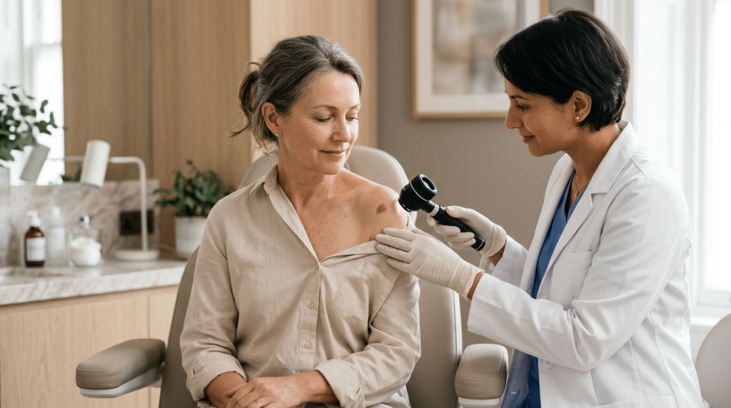
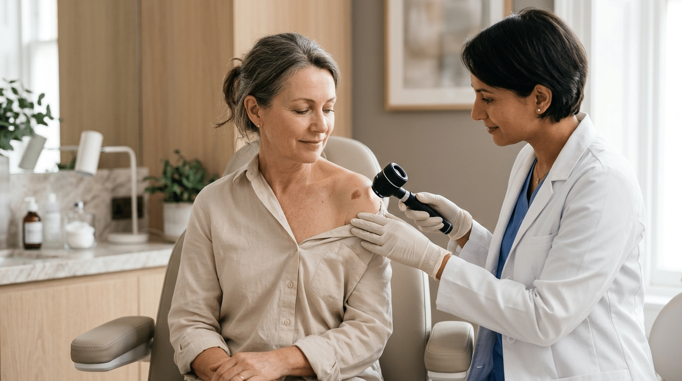
At a specialist assessment, a clinician does considerably more than glance at the growth. Using a dermatoscope — a handheld device that magnifies and illuminates the skin — they can see structures invisible to the naked eye. Seborrhoeic keratoses have characteristic dermoscopic features, such as small keratin-filled cysts and comedo-like openings, that help confirm the diagnosis. Melanomas show a different set of features, such as an atypical pigment network or irregular vascular patterns. In the majority of cases this allows a confident distinction.
Where any doubt remains, the definitive answer comes from removing the lesion and sending it for histopathology — laboratory examination of the tissue under a microscope. This is the gold standard, because it examines the actual cells rather than relying on surface appearance. At Centre for Surgery, any lesion of clinical concern is removed with appropriate margins and sent for histopathology so the diagnosis is confirmed rather than assumed, and anything found to be malignant is directed to the appropriate specialist skin cancer pathway. Benign seborrhoeic keratoses being removed purely for cosmetic reasons can often be treated with erbium laser, which gives an excellent cosmetic result — but this is only chosen once the diagnosis is clear.
Having a seborrhoeic keratosis removed
If your growth has been confirmed as a benign seborrhoeic keratosis and you would like it gone — whether for comfort or appearance — it can be removed straightforwardly. Depending on the size, location and pigmentation of the lesion, options include erbium laser ablation, which is particularly good for a near-scarless finish on the face, or surgical techniques where excision and histology are preferred. Most removals are carried out under local anaesthetic as a day-case procedure. You can read the full detail on our seborrhoeic keratosis removal page, and for an overview of how we assess and treat all benign and suspicious growths, see our skin lesion removal hub.
The experience itself is usually quick and well tolerated. After the area is numbed, a single seborrhoeic keratosis typically takes only a few minutes to treat, and most people are back to normal activities the same day. With erbium laser, the treated area is usually flat immediately afterwards, with some mild pinkness that settles over the following days to weeks. Because seborrhoeic keratoses are superficial, well-chosen laser treatment can remove them while leaving the surrounding skin largely untouched — which is what makes it such a good option for visible areas like the face. Where a growth is removed by excision instead, a small dressing and a short healing period apply, and the tissue goes off for confirmation.
Cost depends on how many lesions you want treated, their size and the technique used, so the only accurate figure is one given after an assessment. For any procedure over £1,500, Centre for Surgery offers 0% APR finance through Chrysalis Finance, subject to status, so treatment can be spread over monthly instalments. Your patient coordinator can go through the options at your consultation.
Frequently asked questions
Can a seborrhoeic keratosis become cancerous over time?
No. A seborrhoeic keratosis is benign and does not transform into skin cancer, no matter how long you have had it. The reason to have a changing growth checked is not that the keratosis itself is becoming dangerous, but that an entirely separate melanoma can occasionally resemble or arise near one.
Why has my seborrhoeic keratosis suddenly changed or grown?
Seborrhoeic keratoses can become irritated, inflamed or slightly larger, particularly if they are caught or rubbed, and they can darken. However, because change is also a warning sign for melanoma, a growth that is genuinely changing should be assessed by a professional rather than assumed to be a simple irritation.
Is it normal to suddenly develop lots of seborrhoeic keratoses?
Developing more of them gradually with age is completely normal. A sudden appearance of a large number in a short space of time is less common and is worth mentioning to a doctor, as very occasionally it can be associated with other health matters that are worth ruling out.
Can I tell the difference myself?
You can learn the typical features, and the ABCDE rule is a useful guide, but you cannot reliably diagnose a pigmented growth by eye — and neither can most clinicians without a dermatoscope. If a growth concerns you or has any warning features, the sensible step is a professional assessment rather than self-diagnosis.
Does a seborrhoeic keratosis need to be removed?
Not for medical reasons. Removal is a personal choice made for comfort or cosmetic preference. The exception is when the diagnosis is uncertain, in which case removal with histology may be recommended to confirm exactly what the growth is.
Book a skin lesion assessment at Centre for Surgery
If you have a growth you are unsure about, the reassuring and sensible step is to have it looked at properly. Centre for Surgery is an independent clinic on Baker Street, rated “Good” across all five domains by the Care Quality Commission, England’s independent healthcare regulator. Every lesion is assessed by a GMC-registered specialist, and any growth of clinical concern is removed and sent for histopathology so the diagnosis is confirmed rather than assumed. Our medical content is reviewed by Consultant Plastic Surgeon Dr Spyridon Vlachos (GMC 7522950).
Call: 0207 993 4849
Email: contact@centreforsurgery.com
Address: 95–97 Baker Street, London W1U 6RN
Online: Book a consultation via our contact form below.
[contact-form-7 id=”256″]