

“Is it flat or raised?” is one of the first questions a surgeon asks about a mole — and it matters more than most people realise. The shape of a mole is not just cosmetic; it hints at how deep the mole sits in the skin, which in turn shapes how it should be removed and, occasionally, how carefully it needs to be checked. This guide explains the real difference between flat and raised moles, clears up a common and slightly dangerous myth about which is “easier” to remove, and sets out how each type is treated during mole removal at Centre for Surgery.
There is one point worth making at the very start, because a lot of cosmetic advice online gets it backwards: a flat mole is not automatically the simple, laser-it-off option, and a raised mole is not automatically the difficult one. As you will see, the opposite is often true.
What is a mole?
A mole — known medically as a melanocytic naevus — is a benign skin growth made up of clusters of pigment-producing cells called melanocytes. Normally these cells are spread evenly through the skin; in a mole, they group together to form a visible spot. Most people have between 10 and 40 moles, most appearing in childhood and early adulthood.
Moles vary enormously. They can be flat or raised, smooth or rough, hairy or hairless, and range in colour from pink and tan through to dark brown or black. The vast majority are entirely harmless and stay the same throughout life. A small number change over time, and a very small number can develop into melanoma, the most serious form of skin cancer — which is why the shape and behaviour of a mole, not just its appearance, are what a specialist pays attention to.
Flat moles vs raised moles: what is the difference?
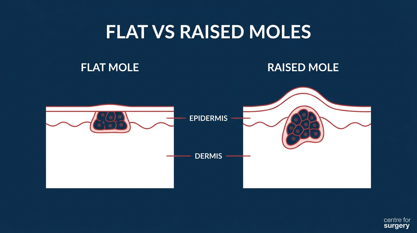
The difference between a flat and a raised mole comes down to where in the skin the pigment cells sit. That single fact explains almost everything else — how the mole looks, how it behaves, and how it is best removed.
Flat moles
Flat moles sit flush with the surface of the skin. You can see them but not really feel them — run a finger over one and the skin feels smooth. In medical terms, most flat moles are junctional naevi: the melanocytes are concentrated at the junction between the epidermis (the top layer of skin) and the dermis (the layer beneath). They are typically round or oval, evenly coloured, and range from light brown to almost black.
Crucially, “flat” does not mean “shallow.” The pigment cells in a flat mole can extend down into the deeper layers of the skin. This is the detail that most cosmetic marketing glosses over, and it has real consequences for removal, which we come to below.
Raised moles
Raised moles sit above the level of the surrounding skin — you can both see and feel them. They may be dome-shaped, smooth, or slightly rough, and are often lighter in colour than flat moles, sometimes skin-toned. Many are compound naevi (pigment cells in both the upper and deeper layers) or dermal naevi (pigment cells mostly in the deeper dermis, with the mole raised well clear of the surface). Raised moles frequently have hair growing through them, which is usually a reassuring sign rather than a worrying one.
Because a raised, benign mole projects above the skin, it can often be removed at surface level without disturbing the deeper layers — which is the key to why raised moles are frequently the simpler ones to remove.
The myth: “flat moles are easier to remove”
A great deal of cosmetic-clinic advice suggests flat moles are the easy ones — ideal for a quick laser or a superficial shave — while raised moles are the tricky ones. In practice, surgeons and dermatologists often find the reverse.
Here is why. A raised, clearly benign mole sits above the skin, so it can be shaved off flush with the surface in a few minutes, no stitches required. A flat mole, by contrast, has its pigment cells sitting at or below the skin surface and sometimes running deep. To remove a flat mole completely — rather than leaving pigment behind that can simply regrow — the surgeon usually has to cut down into the skin and close the wound with stitches. That is a slightly more involved procedure and it leaves a fine linear scar rather than a flat circular mark.
There is a second, more important reason flat moles are treated with more caution. Because flat, pigmented, changing moles are precisely the pattern that can occasionally signal melanoma, a flat mole that has changed is far more likely to warrant complete surgical removal with the tissue sent for laboratory analysis — something a laser cannot allow, because it destroys the very tissue that would need to be examined. So the “easy laser job” framing is not just wrong about difficulty; in the wrong hands it can be unsafe.
How is each type of mole removed?
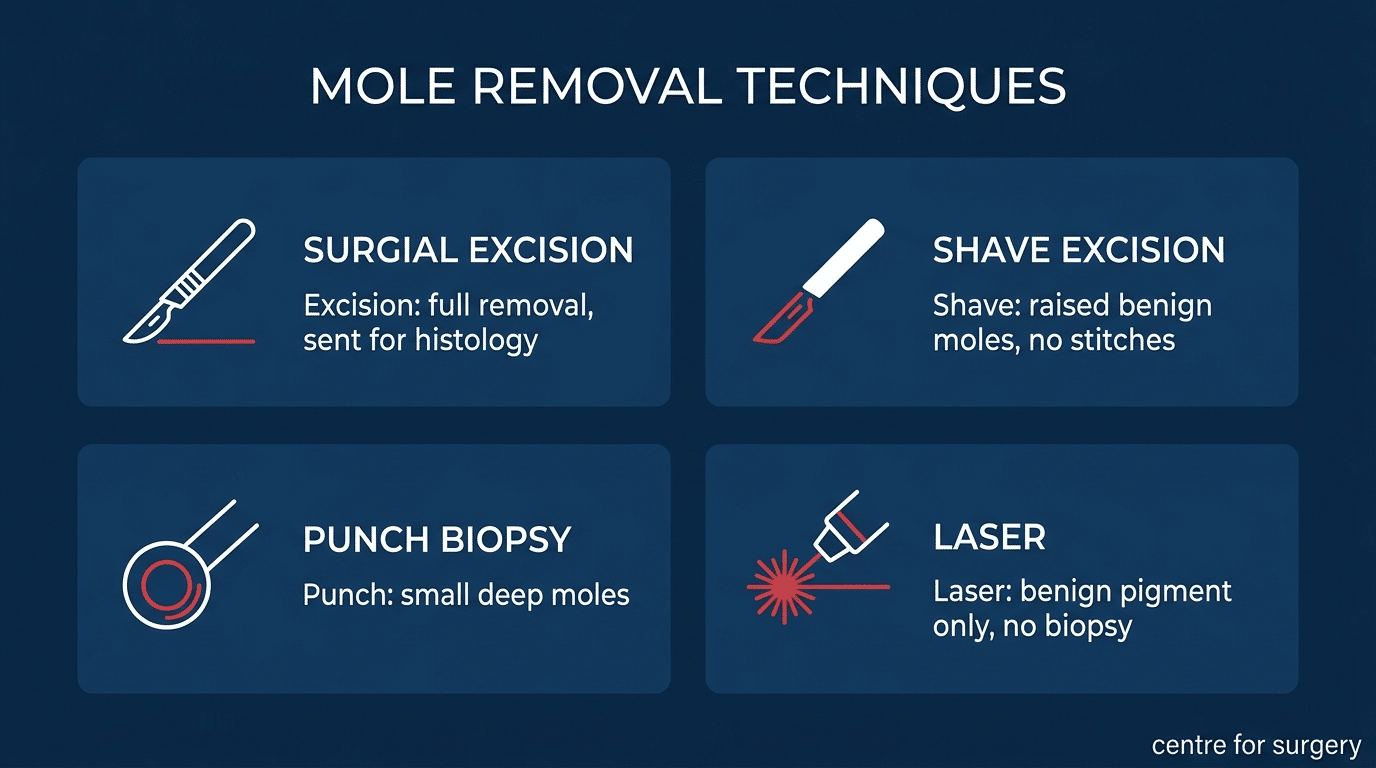
At Centre for Surgery, the removal method is matched to the individual mole — its type, depth, location, and whether there is any clinical concern — rather than forcing every mole through a single technique. There are four main approaches.
Surgical excision
The mole and a small margin of normal-looking skin are removed in one piece, and the wound is closed with sutures. The specimen is sent to an accredited histopathology laboratory and reported by a consultant histopathologist, usually within 7 to 14 days — we explain why in should every removed mole be sent for biopsy. The resulting scar is a fine line, planned along the natural skin tension lines wherever possible, which usually fades to a pale mark over 6 to 12 months. Surgical excision is the method of choice for flat moles, deep moles, and any mole with concerning features, because it removes the whole lesion and allows it to be examined.
Shave excision
Used for raised, benign moles that sit above the surrounding skin. The mole is shaved off flush with a small blade and the base is sealed to control bleeding. No sutures are needed, and the wound heals as a flat circular mark that fades over months. Because the deeper roots of the mole can remain, a small proportion of shave-excised moles regrow over time — uncommon, but worth knowing, and one reason it is reserved for moles that are clearly benign. It is not suitable for flat moles, deep moles, or any mole with concerning features.
Punch biopsy
A circular cutting tool removes a small, deep mole in one core, with the smallest possible diameter of skin. It suits small moles that are too deep for a shave but small enough not to need a full ellipse excision, and the tissue can still be sent for analysis.
Laser
Laser can fade certain small, superficial, clearly benign pigmented marks without cutting the skin. Its major limitation is that it destroys the mole rather than removing it intact, so nothing can be sent to the laboratory. For this reason we never use laser on a mole of any clinical concern, and never on a mole that has changed. Laser has a narrow, genuinely cosmetic role, and only after a specialist has confirmed the mole is harmless — see can all moles be safely removed?
Matching the method to the mole
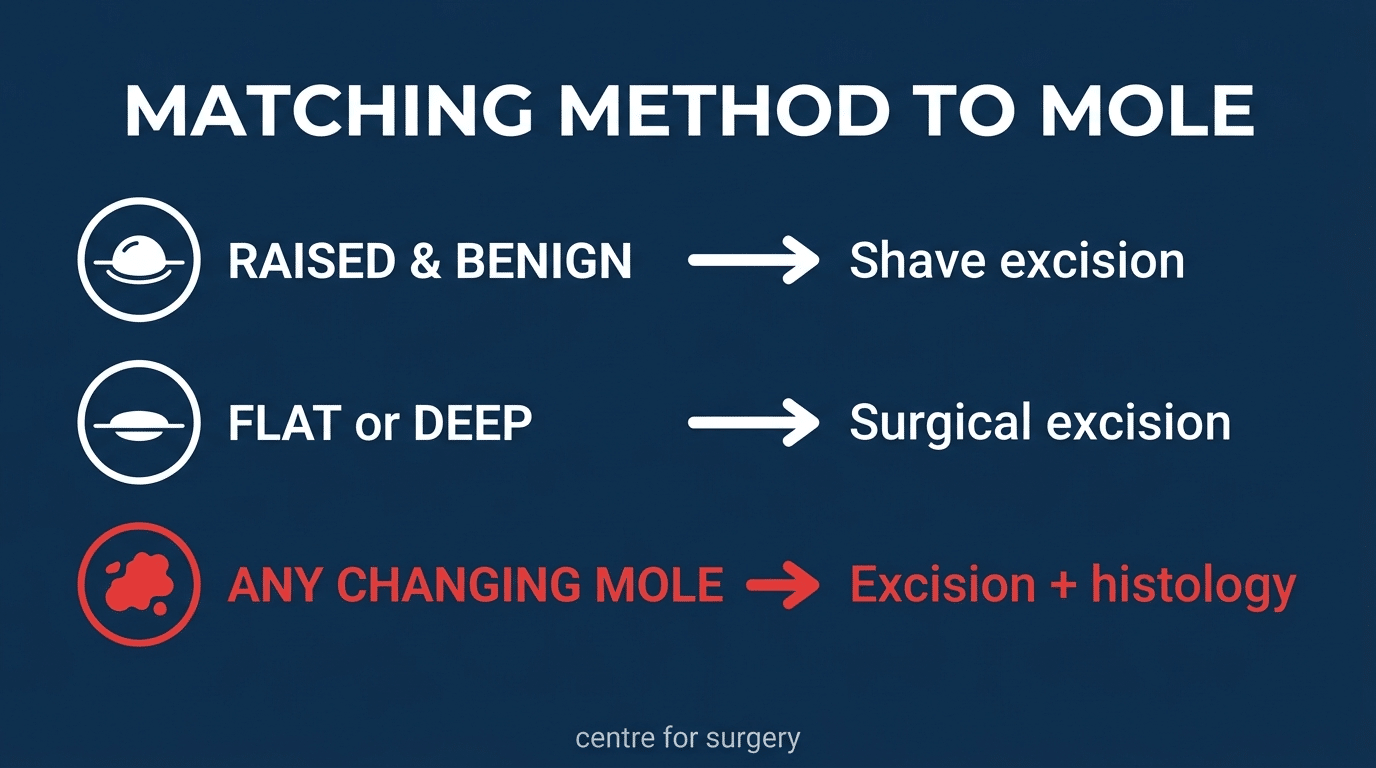
The logic that ties all of this together is straightforward:
- Raised and benign — usually shave excision. Quick, no stitches, minimal mark.
- Flat, or deep — usually surgical excision, because the whole depth of the mole needs to come out and the wound needs closing.
- Any changing or suspicious mole, flat or raised — surgical excision with the tissue sent for histology, never laser. This is the safety rule that overrides all cosmetic considerations.
This is also why being offered only one removal technique is a poor sign. A clinic that only lasers, or only shaves, will inevitably push some moles through the wrong method. A proper assessment starts with the mole, not the machine — which is why choosing a plastic surgeon for mole removal matters.
When should a mole be checked?
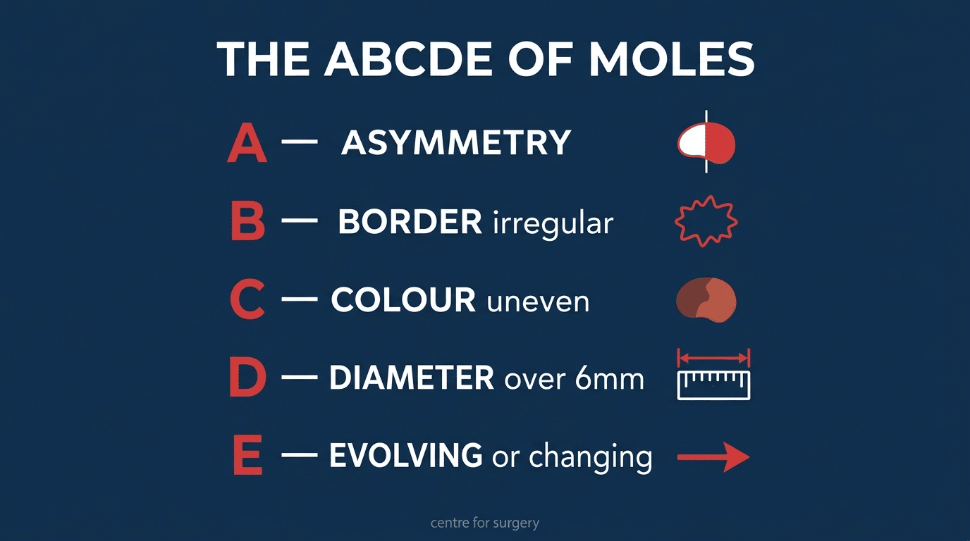
Most moles never need treatment. But some changes are worth acting on, and the widely used ABCDE guide is a helpful memory aid for what to look for:
- A — Asymmetry: one half of the mole does not match the other.
- B — Border: edges that are irregular, ragged, notched, or blurred.
- C — Colour: more than one colour, or uneven shades of brown, black, red, or blue.
- D — Diameter: larger than about 6mm (the size of a pencil-top rubber), though melanomas can be smaller.
- E — Evolving: any change in size, shape, colour, or elevation, or new symptoms such as itching, bleeding, or crusting.
The single most important letter is E. A mole that is changing — whatever it looks like — is the one to have assessed. A mole that has itched, bled, or crusted without an obvious cause such as catching it on clothing should also be checked. If you notice any of these, arrange to see a GP or a specialist promptly; the great majority turn out to be harmless, but it is always worth confirming. You can read more in our guide on whether to be concerned about an itchy or bleeding mole.
Will removing a mole leave a scar?
Any mole removal leaves some mark, but its appearance depends on the method, the mole’s size and location, your skin type, and how you heal. Shave excision tends to leave a flat, circular mark that blends over months. Surgical excision leaves a fine line, planned along the skin’s natural creases where possible, that usually settles to a pale mark over 6 to 12 months. Areas such as the chest, back, and shoulders are more prone to raised or thickened scars in some people, and your surgeon will discuss the likely cosmetic outcome before treatment — particularly for a mole being removed purely for appearance. Our guides on what mole removal scars look like and how long a mole removal scar takes to fade cover this in detail.
Should every removed mole be tested?
Where there is any clinical concern, the answer is an unequivocal yes — the mole is excised whole and sent for histopathology, and this is non-negotiable. For a mole that a specialist is confident is entirely benign and is being removed purely for cosmetic reasons, testing may not be strictly necessary, but many surgeons still send the specimen for peace of mind where the removal method allows it. This is one of the strongest arguments against laser or any method that destroys the tissue: it removes the option of testing altogether. We look at this in more depth in should every removed mole be sent for biopsy.
Flat and raised moles: frequently asked questions
Are raised moles more dangerous than flat moles?
No — shape alone does not indicate danger. Many raised moles are entirely benign dermal naevi, and many flat moles are harmless junctional naevi. What matters is not whether a mole is flat or raised, but whether it is changing. A changing mole of either type should be assessed.
Can a flat mole become raised?
Moles can slowly evolve over years as the balance of pigment cells shifts deeper into the skin — a junctional naevus can gradually become a compound and then a dermal naevus, becoming more raised in the process. Slow change over years is usually normal; relatively rapid change over weeks or months is what warrants a check.
Can flat moles be removed with laser?
Sometimes, but with caution. Laser can only fade a small, superficial, confirmed-benign flat mole, and it prevents any tissue being tested. Because flat moles can sit deeper than they appear and are the pattern more likely to need testing, surgical excision is usually the safer choice. Laser is never appropriate for a flat mole that has changed.
Why does removing a flat mole leave a bigger scar than a raised one?
Because the surgeon usually has to cut down into the skin and close the wound with stitches to remove a flat mole completely, whereas a raised benign mole can be shaved off at the surface. The trade-off is that complete excision removes the whole mole and allows it to be tested.
Do moles grow back after removal?
Fully excised moles do not usually return. Shave-excised moles can occasionally regrow if some pigment cells remain in the deeper skin, which is why suspicious moles are always fully excised rather than shaved.
How many moles can be removed at once?
Several moles can often be removed in a single visit, depending on their size and location. Your surgeon will advise at consultation what is sensible to do in one sitting. Moles are one of several common lumps and bumps we remove.
Does mole removal hurt?
The procedure is carried out under local anaesthetic, so you should feel only the initial small injection and then pressure rather than pain. Most moles are removed in a few minutes.
Mole removal at Centre for Surgery
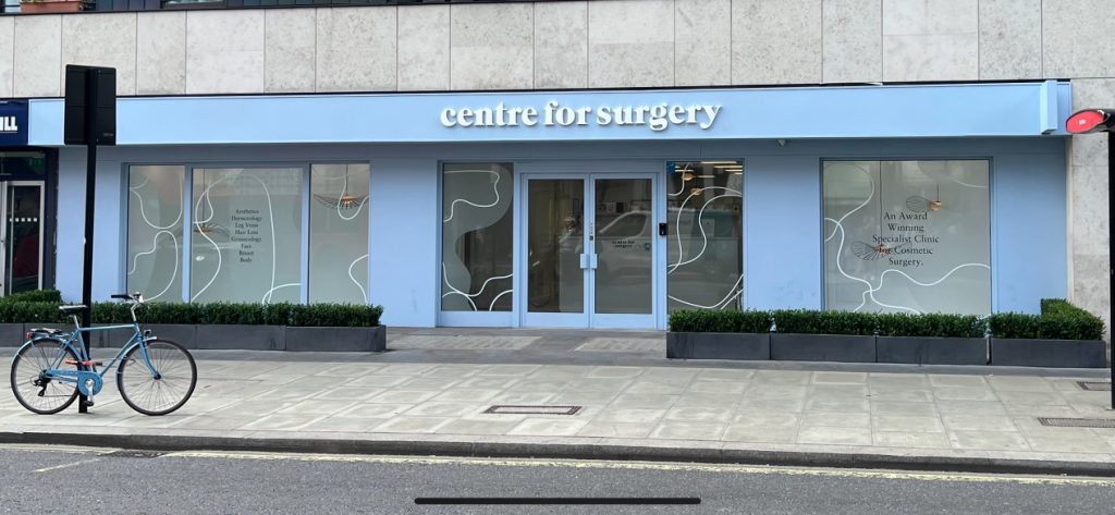
Centre for Surgery is an independent clinic at 95–97 Baker Street in London, rated “Good” across all five domains by the Care Quality Commission. Our medical content is reviewed by Consultant Plastic Surgeon Dr Spyridon Vlachos (GMC 7522950).
Every mole removal at Centre for Surgery is preceded by a consultation with a GMC-registered specialist who can offer all four techniques — surgical excision, shave, punch, and laser — and choose the most appropriate one for your particular mole. Where any feature suggests possible skin cancer, we excise the mole and send it for histopathology; we never use laser to ablate a mole of clinical concern. A few things set our approach apart:
- Consultation with your surgeon — the person assessing your mole is the person who removes it.
- All four techniques available — the method is matched to your mole, not to the single machine a clinic happens to own.
- Histology as standard where indicated — reported by a consultant histopathologist within 7 to 14 days.
- Scar-conscious technique — incisions planned along natural skin tension lines, with aftercare guidance to help scars settle.
- A CQC-regulated clinic — with the follow-up and oversight that comes with it.
To have a mole assessed or removed, book a consultation, call 0207 993 4849, or email contact@centreforsurgery.com to arrange an in-person consultation at our Baker Street clinic.
For procedures over £1,500, 0% APR finance is available through Chrysalis Finance, subject to status. See our finance options for details.
Related Articles
- Should I Be Concerned About an Itchy or Bleeding Mole?
- What Do Mole Removal Scars Look Like?
- Can I Get Mole Removal on the NHS?
[contact-form-7 id=”256″ title=”Treatments form”]